

Device Lab 68
X-ray
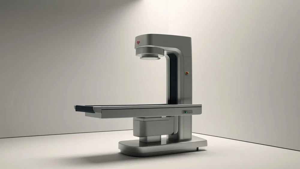
A floor-mounted tube column on a rail, a high-frequency generator in a cabinet, a table with a floating top and a Bucky tray under it, an erect stand against the wall, and a control console behind lead glass. In the tray, in front of the digital detector, sit a twelve-to-one focused grid and three ionisation chambers.
Also called: X-Ray Machine, Radiography, X Ray. Arabic: جهاز الأشعة السينية. Device Lab 68 of 69 in BMEssentia Medical City, 2 configurations.
Overview: the configurations
Fixed radiographic room, table Bucky with grid and automatic exposure control
A floor-mounted tube column on a rail, a high-frequency generator in a cabinet, a table with a floating top and a Bucky tray under it, an erect stand against the wall, and a control console behind lead glass. In the tray, in front of the digital detector, sit a twelve-to-one focused grid and three ionisation chambers.
Mobile ward unit, no grid and no automatic exposure control
A trolley with a battery store, a column with a boom arm, the tube and collimator at the end of it, a console on the body and a flat detector that is carried to the patient and slid behind them. There is no grid and no chamber. The exposure is set from a chart on the machine and a judgement of how thick the patient is, and then it happens.
How it works: the main parts
What each part does, from the X-ray lessons in the 3D Device Lab.
- Cathode filaments and focusing cup
- Two coils of tungsten wire, each a few millimetres long, lying in slots cut into a block of nickel at the far end of the tube from the target. One coil is short and one is longer.
- Rotating anode, target track and rotor
- A disc of molybdenum about ninety millimetres across with a band of tungsten and rhenium brazed onto a bevel at its rim, spinning at several thousand revolutions a minute inside the vacuum. The bevel is the most important angle in the device.
- Tube housing, insulating oil and lead shielding
- A heavy cylinder, lead-lined, filled with oil, with the tube insert suspended inside it and one square window in the wall where the beam is allowed out. It weighs far more than the tube it contains, and almost all of that weight is doing two jobs: holding off a hundred kilovolts and absorbing the photons that were not asked for.
- Tube port and inherent filtration
- A thin window in the envelope where the beam leaves, the oil in front of it, and the window in the housing wall beyond that. Together they are the filtration nobody chose.
- Added aluminium filtration
- Two thin sheets of aluminium in a drawer under the port, held by a clip, with a handle to pull them out. They are the cheapest and least interesting-looking part of the machine, and removing them raises the exposure a patient absorbs without improving the radiograph at all.
- Collimator, light field and lead shutters
- A box bolted under the port with two pairs of lead shutters inside it, a lamp, a mirror at forty-five degrees and a pair of cross-hair wires. The lamp and the mirror exist so that somebody can see where an invisible beam is going.
- High-frequency generator and high-tension transformer
- A cabinet the size of a wardrobe holding an inverter, a transformer in oil with two bushings on top, a rectifier stack and a control board. It decides how fast the electrons are going when they hit the target, and that single decision is what makes bone look different from soft tissue.
- High-tension cables and receptacles
- Two cables as thick as a wrist, running from the generator to the two ends of the tube housing, each ending in a plug that screws into a well full of insulating grease. They are the least glamorous expensive part of the room and the one most often damaged by the room being rearranged.
Explore it in the Device Lab
- The exchange rate between the two knobs: Hold the receptor exposure where it is and raise the kilovoltage by fifteen per cent, taking out the milliampere-seconds that the extra penetration allows. Watch three numbers: what the receptor gets, what the patient absorbs, and the contrast.
- Where the contrast actually comes from: Sweep the kilovoltage and watch one quantity that is not on any real console: the fraction of cortical bone’s attenuation that is NOT Compton scattering.
- Two ways to deal with scatter, and only one is free: Set a thick patient and a wide field, then reduce the field and watch the scatter-to-primary ratio fall. Then restore the field and put a grid in instead, and watch the contrast recover at the price of a large increase in exposure.
- Why the fixed system is the blurrier one: Compare the penumbra on the two configurations. 15 m and the mobile unit at one metre, so the fixed room should be the sharper of the two. It is not.
- Measuring it, or predicting it: Make the patient six centimetres thicker on each configuration in turn and watch what happens.
Common faults and what to check
Most reported device problems (FDA MAUDE)
| Device problem | Reports |
|---|---|
| Device Fell | 264 |
| Device Handling Problem | 247 |
| Image Display Error/Artifact | 229 |
| Device Operational Issue | 153 |
| Break | 152 |
| Component Falling | 134 |
Raw counts of adverse event reports for this device type (FDA product code KPR, MQB), all years. Not adjusted for the number of units in use, so they show what gets reported, not failure rates. Report text is never copied.
Planned maintenance checks
From the BMEssentia procedure library: every 12 months, about 120 minutes, electrical class I, applied part type B.
| Check | Pass when |
|---|---|
| Tube stand, cables and room Inspect the tube stand locks, the high-voltage cables, the table and bucky, the warning lights and the lead aprons. | Locks hold; no cable damage; warning lights work. |
| Electrical safety Test the table and mobile parts to IEC 62353. | PE ≤ 0.30 Ω; equipment leakage ≤ 500 µA (Class I). |
| kVp, time, reproducibility and linearity With the meter on the table, expose at 60, 80 and 100 kVp; repeat one setting five times; step through adjacent mA settings. | kVp and time within the manufacturer's accuracy; CV ≤ 0.05; linearity within 0.10 of the sum. |
| Half-value layer At 80 kVp add aluminium filters until the dose halves. | At least the regulatory minimum for 80 kVp. |
| Collimation and alignment Expose the collimation tool and compare the light field with the X-ray field. | Total misalignment per axis within 2% of SID. |
Test instruments: Non-invasive kVp and dose meter (multimeter for X-ray); Aluminium filters for HVL; Collimation test tool; Electrical safety analyser.
Standards engineers test against
IEC 60601-2-54: radiography and radioscopy (X-ray)
The particular standard for X-ray equipment for radiography and fluoroscopy.
Recent FDA recalls
| Recall | Product and reason |
|---|---|
| Z-2354-2026 2026-06-05 | Philips North America Llc: DigitalDiagnost C90 Flex/Value/Chest/ER. Model Number: 712035. System was delivered without the required Seismic kit for Wall Stand VS2, which is designed to prevent damage and ensure stability of the Wall Stand VS2 during seismic events. Root cause: Device Design. |
| Z-1299-2026 2026-02-05 | FUJIFILM Healthcare Americas Corporation: FDR Visionary Suite It was found that the contacts of the electromagnetic contactors used inside the cabinet of the X-ray high-voltage generator were welded, and the current might continue to be supplied to the power supply circuit even if. Root cause: Process control. |
| Z-0464-2026 2025-11-07 | Philips Medical Systems DMC GmbH: DigitalDiagnost 4 (1) High Performance, (2) Flex/Value Philips has identified that some ceiling suspension telescopic carriages manufactured from July 2024 through June 2025 may be difficult to move vertically (i.e. Root cause: Device Design. |
| Z-0466-2026 2025-11-07 | Philips Medical Systems DMC GmbH: Radiography 7300 C Philips has identified that some ceiling suspension telescopic carriages manufactured from July 2024 through June 2025 may be difficult to move vertically (i.e. Root cause: Device Design. |
| Z-0465-2026 2025-11-07 | Philips Medical Systems DMC GmbH: DigitalDiagnost C90 (1) High Performance, (2) Flex/Value/Chest/ER Philips has identified that some ceiling suspension telescopic carriages manufactured from July 2024 through June 2025 may be difficult to move vertically (i.e. Root cause: Device Design. |
From the FDA recall database (openFDA), newest first, imported 2026-10-22. 363 recalls on record for this device type. Recall text is the FDA's public text; each recall links to the official record.
Learn it, see it in 3D, ask about it
- Ask Essentia about the X-ray: answers from named sources, with the device open in its 3D Device Lab beside the answer.
- The X-ray on the BMEssentia Platform: the Device Library course, 13 sections from physics to maintenance.
- BMEssentia Medical City: Device Lab 68, the X-ray fully assembled in 3D, with lab tasks and the simulations.
Previous: TENS. Next: Lithotripter (ESWL). All 69 devices.
Ask about the X-ray
Sign in with your BMEssentia account, or create one, and ask anything about this device.
Ask Essentia