

Essentia / Devices / Ultrasound
Device Lab 36
Ultrasound
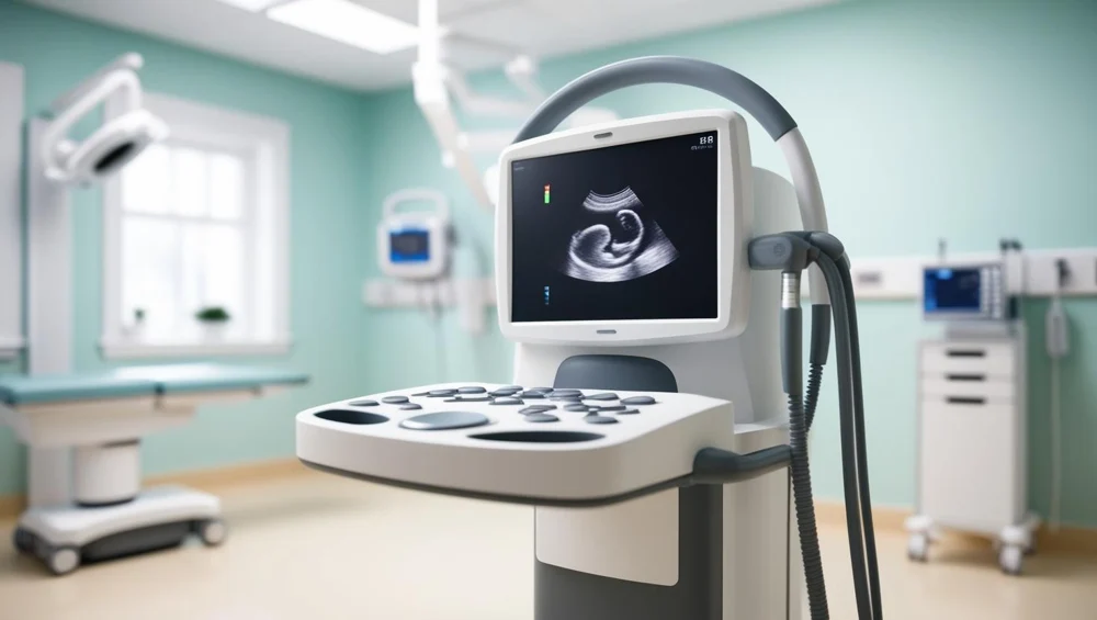
A flat array of a hundred and twenty-eight elements diced at about three tenths of a millimetre, running at ten megahertz. It does not steer. It fires a group of adjacent elements straight down, forms one line, then slides the active group one element along and does it again, which is why the image is a rectangle exactly as wide as the probe.
Also called: Ultrasound Machine, Ultrasound Scanner, Sonography. Arabic: جهاز الموجات فوق الصوتية. Device Lab 36 of 69 in BMEssentia Medical City, 2 configurations.
Overview: the configurations
Linear array, vascular and superficial
A flat array of a hundred and twenty-eight elements diced at about three tenths of a millimetre, running at ten megahertz. It does not steer. It fires a group of adjacent elements straight down, forms one line, then slides the active group one element along and does it again, which is why the image is a rectangle exactly as wide as the probe.
Phased array, cardiac and deep
Sixty-four elements diced at almost exactly the same physical pitch as the linear probe, in a footprint a third of the width, running at two and a half megahertz. Every element fires on every line, with a delay across the array that tilts the wavefront, so the beam sweeps out a sector from what is very nearly a point.
How it works: the main parts
What each part does, from the Ultrasound lessons in the 3D Device Lab.
- Mains inlet, isolation and protective earth
- An inlet, a fuse and an earth stud on the back of the tower, and behind them something most equipment in the building does not have: a transformer whose only job is to make sure the patient is not connected to the mains by any route at all.
- Transmit pulser
- A bank of high-voltage switches, one per channel, that slam a hundred volts or so across a piezoelectric element for a fraction of a microsecond and then stop. It is the loudest thing in the machine electrically and it does nothing at all for the other ninety-nine per cent of the time.
- Transmit and receive beamformer
- The board with the largest chip on it and the most channels running to it. Nothing about it moves and nothing about it is acoustic, and it is where the beam actually comes from.
- Preamplifier and time-gain compensation
- A row of small amplifiers immediately behind the connector, one per channel, and a gain that climbs while it is listening. It is the first thing the echo meets and the last chance to amplify it before noise is added.
- Digitiser
- A row of converters turning each channel into numbers, tens of millions of times a second. Everything before it is analogue and fragile; everything after it is arithmetic and can be copied without loss.
- Envelope detection, compression and scan conversion
- The stage that turns a line of radiofrequency signal into a line of grey pixels, and then turns a fan of such lines into a rectangle a screen can display. Three operations, each of which throws information away on purpose.
- Upper display
- The large screen on the articulated arm, angled towards whoever is reading rather than whoever is scanning. In a room with the lights down it is the brightest object in the space.
- Touchscreen control display
- The smaller angled screen above the keys, raked back so a seated operator can reach it without looking away from the patient for long. Its content changes with the mode, which is the whole reason it exists.
Explore it in the Device Lab
- The account everything is bought from: Change the frequency and watch four numbers at once: axial resolution, lateral resolution, penetration and the mechanical index.
- The number that decides whether a probe can steer: Set the steering angle to zero and step it outwards, watching for the angle at which a grating lobe appears. Do it on both probes.
- One number that is three things: Change the depth and watch the pulse repetition frequency, the frame rate and the Nyquist velocity all move together.
- Why bone is white and air is a wall: Compute the reflection coefficient at three boundaries: tissue against fat, tissue against bone and tissue against air.
- What 1540 metres per second costs: Set a speed-of-sound error and watch the reported depth against the real one.
Common faults and what to check
Most reported device problems (FDA MAUDE)
| Device problem | Reports |
|---|---|
| Poor Quality Image | 3,118 |
| Retraction Problem | 1,037 |
| Break | 587 |
| Defective Component | 573 |
| Device Displays Incorrect Message | 373 |
| Battery Problem | 361 |
Raw counts of adverse event reports for this device type (FDA product code IYO, ITX), all years. Not adjusted for the number of units in use, so they show what gets reported, not failure rates. Report text is never copied.
Planned maintenance checks
From the BMEssentia procedure library: every 12 months, about 60 minutes, electrical class I, applied part type BF.
| Check | Pass when |
|---|---|
| Probes and cables Inspect every probe lens for cracks, delamination and bubbles, and the cables and strain reliefs. | No cracks or delamination; cable intact. |
| Electrical safety Test the scanner to IEC 62353 and leakage-test the endocavity and TOE probes. | PE ≤ 0.30 Ω; equipment leakage ≤ 500 µA; applied part BF ≤ 5000 µA (or CF ≤ 50 µA if so rated). |
| In-air reverberation With the probe in air, view the reverberation pattern at the default preset. | Even pattern across the face; no dark vertical bands (dead elements). |
| Phantom image quality Scan the phantom with each probe at a fixed preset and compare dead zone, resolution and depth of penetration with the baseline. | Within the baseline. |
Test instruments: Tissue-mimicking QA phantom; Electrical safety analyser; Probe leakage tester (for endocavity and TOE probes).
Standards engineers test against
IEC 60601-2-37: ultrasonic diagnostic and monitoring equipment
The particular standard for diagnostic ultrasound.
Recent FDA recalls
| Recall | Product and reason |
|---|---|
| Z-3276-2026 2026-09-25 | Volcano Corp: Philips IntraSight Plus Software system may associate image data from one patient with another patient's record, potentially resulting in incorrect diagnosis or treatment. Root cause: Software Design Change. |
| Z-1731-2026 2026-04-01 | Civco Medical Instruments Co. Inc.: eTRAX Needle System Starter Kit 12G (for Aurora Trackers), Part Number 667-149 There was an error in inspection and programming of the eTRAX needle sensor for Aurora trackers. Root cause: Software change control. |
| Z-1732-2026 2026-04-01 | Civco Medical Instruments Co. Inc.: eTRAX Needle System Starter Kit 14G (for Aurora Trackers), Part Number 667-150 There was an error in inspection and programming of the eTRAX needle sensor for Aurora trackers. Root cause: Software change control. |
| Z-1733-2026 2026-04-01 | Civco Medical Instruments Co. Inc.: eTRAX Needle System Starter Kit 16G(for Aurora Trackers), Part Number 667-151 There was an error in inspection and programming of the eTRAX needle sensor for Aurora trackers. Root cause: Software change control. |
| Z-1734-2026 2026-04-01 | Civco Medical Instruments Co. Inc.: eTRAX Needle System Starter Kit 18G (for Aurora Trackers), Part Number 667-152 There was an error in inspection and programming of the eTRAX needle sensor for Aurora trackers. Root cause: Software change control. |
From the FDA recall database (openFDA), newest first, imported 2026-10-22. 230 recalls on record for this device type. Recall text is the FDA's public text; each recall links to the official record.
Learn it, see it in 3D, ask about it
- Ask Essentia about the Ultrasound: answers from named sources, with the device open in its 3D Device Lab beside the answer.
- The Ultrasound on the BMEssentia Platform: the Device Library course, 13 sections from physics to maintenance.
- BMEssentia Medical City: Device Lab 36, the Ultrasound fully assembled in 3D, with lab tasks and the simulations.
Previous: Colonoscope. Next: Transcranial Doppler. All 69 devices.
Ask about the Ultrasound
Sign in with your BMEssentia account, or create one, and ask anything about this device.
Ask Essentia