

Essentia / Devices / Transcranial Doppler
Device Lab 37
Transcranial Doppler
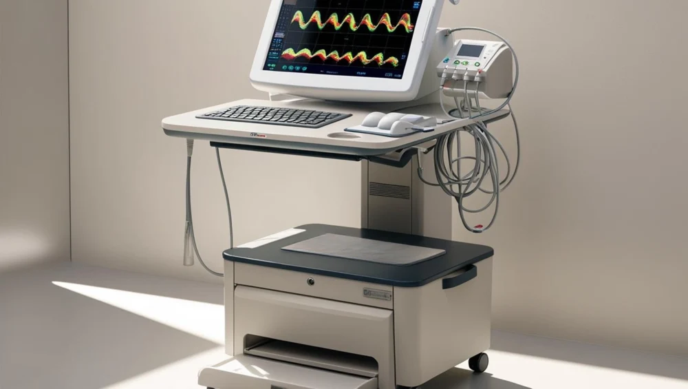
One two-megahertz element about ten millimetres across, in a moulded grip with an orientation flat on it, held against the side of a head by a person who is listening. There is no image of any kind. What the operator has is a depth, a direction, a spectrum and a sound, and finding the vessel means sweeping the probe across the window until the sound is right and then holding still.
Also called: Transcranial Doppler Ultrasound (TCD). Arabic: دوبلر عبر الجمجمة. Device Lab 37 of 69 in BMEssentia Medical City, 2 configurations.
Overview: the configurations
Hand-held non-imaging probe, positioned by the operator
One two-megahertz element about ten millimetres across, in a moulded grip with an orientation flat on it, held against the side of a head by a person who is listening. There is no image of any kind. What the operator has is a depth, a direction, a spectrum and a sound, and finding the vessel means sweeping the probe across the window until the sound is right and then holding still.
Head-frame mounted bilateral probes for continuous monitoring
Two probes, each about eight millimetres across, carried in ball-and-socket gimbals on a frame that clamps round the head with an occipital strap and two tension screws. The gimbals are aimed once, by hand, listening, and then locked, and after that nobody touches them. The frame exists for one reason: so that the angle in the Doppler equation, whatever it is, is the same angle tomorrow morning as it was tonight.
How it works: the main parts
What each part does, from the Transcranial Doppler lessons in the 3D Device Lab.
- Mains inlet, isolation and protective earth
- A filtered appliance inlet, a fuse, an equipotential stud and a rating plate on the back of a box the size of a shoebox, and behind them a transformer whose entire job is to make certain that no single failure anywhere in this machine can put a voltage onto a metal-shelled probe that somebody is pressing firmly against a patient's head.
- Transmit pulser and burst gate
- A high-voltage switch that slams a few tens of volts across a piece of ceramic, a counter that decides how many cycles of that to allow through, and a protection switch that has to disconnect a microvolt amplifier from all of it and reconnect it a few microseconds later.
- Two megahertz element and backing block
- A disc of piezoelectric ceramic about a centimetre across and a millimetre thick, with a heavy lossy block cemented to the back of it, and between them they decide nearly everything about what this device can and cannot reach.
- Quarter-wavelength matching layer and lens
- A layer tens of micrometres thick between the ceramic and the outside world, and a moulded silicone face over the top of it, and between them they are the difference between a probe that works and a probe that reflects nearly everything back into itself.
- Coupling gel and the acoustic window path
- A bottle of warmed gel in a clip on the trolley and a film of it a millimetre thick between a lens and a scalp, and it is not an accessory. It is the first component of the acoustic path and the commonest reason the path stops existing.
- Probe cable and connector
- A metre or two of screened coaxial cable with a multi-pin connector on the end, carrying tens of volts one way and a few microvolts the other way down the same conductor, a few microseconds apart.
- Preamplifier and depth-dependent gain
- A low-noise first stage under a shielding can, followed by an amplifier whose gain climbs as the echo gets later, because a later echo has travelled further and lost more.
- Quadrature demodulator and direction resolution
- Two mixers, a phase splitter that feeds them the same reference ninety degrees apart, and the reason this device knows whether blood is coming towards the probe or going away from it. One mixer on its own could never tell.
Explore it in the Device Lab
- The cosine nobody measured: Set the angle to zero and step it outwards, watching four numbers at once: the velocity, the pulsatility index, the per-degree sensitivity and the displayed wrap limit.
- What the unknown cannot touch: Set a large angle, forty degrees, which under-reads every velocity by twenty-three per cent, and compare the pulsatility index the machine computes against the index of the waveform it was computed from.
- Two products and no free direction: Raise the pulse repetition frequency and watch the wrap velocity rise and the unambiguous depth fall. Then multiply them together.
- Where the budget goes: Compute the round-trip loss through the bone and through the brain separately, and compare them.
- The gate, and what a longer one costs: Lengthen the gate and watch the signal rise and the axial extent of the sample volume rise with it.
Common faults and what to check
Most reported device problems (FDA MAUDE)
| Device problem | Reports |
|---|---|
| Unintended System Motion | 532 |
| Application Program Freezes or Fails to Launch | 158 |
| Material Integrity Problem | 140 |
| Electrical /Electronic Property Problem | 95 |
| Poor Quality Image | 88 |
| Loss of Data | 87 |
Raw counts of adverse event reports for this device type (FDA product code IYN), all years. Not adjusted for the number of units in use, so they show what gets reported, not failure rates. Report text is never copied.
Planned maintenance checks
From the BMEssentia procedure library: every 12 months, about 30 minutes, electrical class I, applied part type BF.
| Check | Pass when |
|---|---|
| Probes and headframe Inspect the probe faces, cables and the headframe. | No cracks. |
| Electrical safety Test to IEC 62353. | Within limits. |
| Velocity check Measure the phantom's known velocity. | Within accuracy. |
Test instruments: Doppler flow phantom (if available); Electrical safety analyser.
Recent FDA recalls
| Recall | Product and reason |
|---|---|
| Z-0152-2027 2026-10-16 | Philips Ultrasound, LLC: Philips Lumify Diagnostic Ultrasound System Ultrasound system may: 1) Not recognize previous transducer registration, prevent scanning, require re-registration with Wi-Fi/cellular access, which may be unavailable outside of the hospital. Root cause: Device Design. |
| Z-3249-2026 2026-09-18 | Philips Ultrasound, LLC: Philips EPIQ Elite Diagnostic Ultrasound System Due to software issue with ultrasound system, doppler sample is displayed shallower than its actual measurement location, potentially causing inaccurate blood flow velocity measurements and misdiagnosis. Root cause: Software Design Change. |
| Z-1007-2026 2025-12-31 | Philips Ultrasound, LLC: Philips EPIQ Ultrasound Systems (various models) used in conjunction with the X5-1c transducer Retroactive: Ultrasound may experience an unexpected automatic reboot, resulting in damage to transducer. Root cause: Software Design Change. |
| Z-0883-2026 2025-12-04 | Philips Ultrasound, LLC: Lumify Diagnostic Ultrasound System models : 795005, 795161, 989605449841, Ultrasound system compatibility issues with Apple devices running iOS 18 may cause a failure to perform live imagining. Root cause: Software design. |
| Z-0435-2026 2025-11-06 | B-K Medical A/S: 1202 Flex Focus Ultrasound System, cart models UA1214 and UA1814, used with: Flex Focus 200, Flex Focus 300, Flex Focus GE HealthCare has become aware that the Instructions for Use within the user guide and service manual do not adequately state the required battery replacement intervals, end-of-life handling procedures, or general. Root cause: Device Design. |
From the FDA recall database (openFDA), newest first, imported 2026-10-22. 300 recalls on record for this device type. Recall text is the FDA's public text; each recall links to the official record.
Learn it, see it in 3D, ask about it
- Ask Essentia about the Transcranial Doppler: answers from named sources, with the device open in its 3D Device Lab beside the answer.
- The Transcranial Doppler on the BMEssentia Platform: the Device Library course, 13 sections from physics to maintenance.
- BMEssentia Medical City: Device Lab 37, the Transcranial Doppler fully assembled in 3D, with lab tasks and the simulations.
Previous: Ultrasound. Next: Cardiac Monitor. All 69 devices.
Ask about the Transcranial Doppler
Sign in with your BMEssentia account, or create one, and ask anything about this device.
Ask Essentia