

Device Lab 5
SPECT
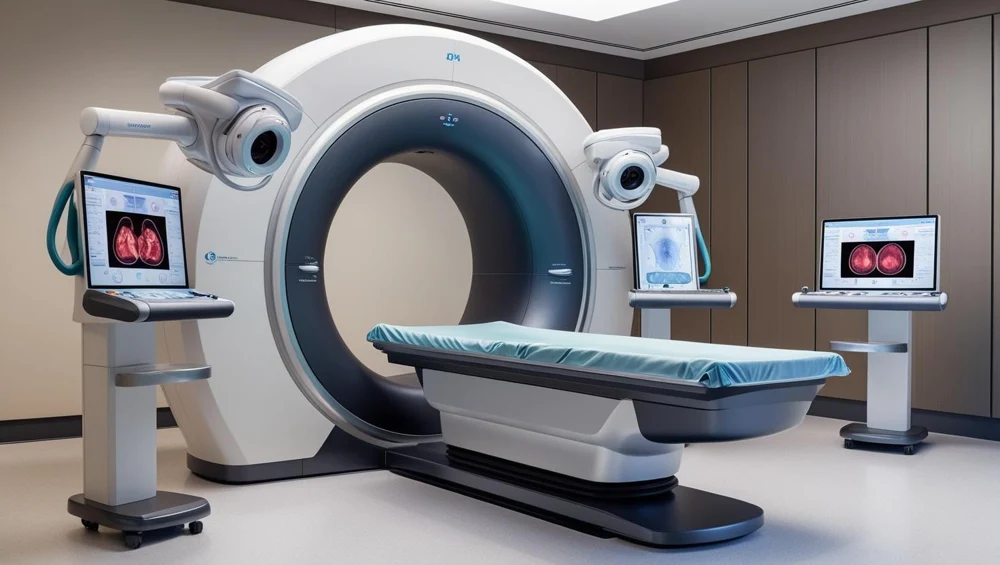
A gamma camera on a rotating gantry. Two detector heads orbit the patient and record where photons left the body, from enough directions to reconstruct a volume.
Also called: Single Photon Emission, Gamma Camera. Arabic: التصوير المقطعي بإصدار الفوتون المفرد. Device Lab 5 of 69 in BMEssentia Medical City, 2 configurations.
Overview: the configurations
Dual-head variable-angle SPECT
A gamma camera on a rotating gantry. Two detector heads orbit the patient and record where photons left the body, from enough directions to reconstruct a volume.
SPECT/CT with attenuation correction
The same gamma camera with a low-dose CT behind it on the same axis. The CT measures how much each path through the patient attenuates, and the reconstruction uses that rather than assuming it.
How it works: the main parts
What each part does, from the SPECT lessons in the 3D Device Lab.
- Collimator
- A slab of lead drilled with tens of thousands of parallel holes, bolted to the face of the detector head. It weighs between eighty and two hundred kilograms, it is changed by hand on a trolley, and it throws away more than ninety-nine point nine per cent of everything that reaches it.
- Scintillation crystal
- A single sheet of sodium iodide about 540 by 400 millimetres and under a centimetre thick, sealed behind a thin aluminium window. It is the point at which a gamma photon stops being radiation and becomes light.
- Light guide
- A slab of optically clear material between the crystal and the photomultipliers, doing something that sounds like a mistake: deliberately spreading the light out.
- Photomultiplier array
- Around fifty tubes in a hexagonal close packing behind the light guide, each one turning a few hundred photons of visible light into a measurable pulse of current.
- Detector head shielding
- The lead that surrounds everything except the collimator face. A detector head weighing four hundred kilograms is mostly this.
- Position and energy electronics
- The boards immediately behind the tubes that turn a set of simultaneous pulses into three numbers: x, y, and how much energy the event deposited.
- Pulse height analyser
- The circuit that looks at each event’s energy and decides whether it is counted or discarded. On a typical technetium study it throws away most of what reaches it.
- Acquisition computer
- The computer that records accepted events and assembles them into projections. On a tomographic study it is also what tells the gantry when to move.
Explore it in the Device Lab
- Resolution and sensitivity are the same dial: Change the collimator and watch two numbers move in opposite directions, every time, without exception. Longer, narrower holes accept a narrower range of directions, so the image resolves better and fewer photons get through. Shorter, wider holes accept more, so the count rate rises and the resolution falls.
- The free control nobody writes down: Resolution at the object falls off with distance from the collimator face, roughly linearly, because the cone of directions a hole accepts widens with distance. Move a head from 250 mm to 150 mm from the axis and the system resolution improves by about a third.
- Energy is a direction test in disguise: The camera measures how much energy each photon had, and it does so to find out whether the position it just recorded is a lie.
- Counts you cannot get back: Set the delay between injection and imaging to six hours and watch what is left. For technetium-99m, with a six-hour half-life, about half. Those counts are not delayed; they are gone, and noise rises as the square root of what remains.
- The quantity the camera cannot measure: Put a source eighty millimetres inside a body of soft tissue and count what comes out. Roughly a third of what was emitted. Put the same source near the surface and almost all of it arrives.
Common faults and what to check
Most reported device problems (FDA MAUDE)
| Device problem | Reports |
|---|---|
| Nonstandard Device | 89 |
| Unintended System Motion | 81 |
| Loose or Intermittent Connection | 74 |
| Component Falling | 73 |
| Device Operates Differently Than Expected | 56 |
| Mechanical Problem | 53 |
Raw counts of adverse event reports for this device type (FDA product code KPS), all years. Not adjusted for the number of units in use, so they show what gets reported, not failure rates. Report text is never copied.
Planned maintenance checks
From the BMEssentia procedure library: every 12 months, about 180 minutes, electrical class I, applied part type B.
| Check | Pass when |
|---|---|
| Heads, collimators and table Inspect the detector heads, collimators (dents), touch sensors and the table. | No collimator damage; touch sensors stop motion. |
| Electrical safety Test to IEC 62353. | Within limits. |
| Uniformity, energy and centre of rotation Acquire floods, check the energy peak and run the centre-of-rotation test. | Within the manufacturer's limits. |
Test instruments: Point source or flood source (per manufacturer); Phantoms for uniformity and resolution; Dose calibrator.
Recent FDA recalls
| Recall | Product and reason |
|---|---|
| Z-0290-2026 2025-10-23 | GE MEDICAL SYSTEMS ISRAEL, FUNCTIONAL IMAGING: Elscint Cardial GE HealthCare has become aware that certain Nuclear Medicine systems that are past the End of Guaranteed Service could have been transported or relocated without adequate detector support. Root cause: Package design/selection. |
| Z-0291-2026 2025-10-23 | GE MEDICAL SYSTEMS ISRAEL, FUNCTIONAL IMAGING: Elscint Model 4XX GE HealthCare has become aware that certain Nuclear Medicine systems that are past the End of Guaranteed Service could have been transported or relocated without adequate detector support. Root cause: Package design/selection. |
| Z-0292-2026 2025-10-23 | GE MEDICAL SYSTEMS ISRAEL, FUNCTIONAL IMAGING: Elscint SPX4 GE HealthCare has become aware that certain Nuclear Medicine systems that are past the End of Guaranteed Service could have been transported or relocated without adequate detector support. Root cause: Package design/selection. |
| Z-0293-2026 2025-10-23 | GE MEDICAL SYSTEMS ISRAEL, FUNCTIONAL IMAGING: Elscint SPX6 GE HealthCare has become aware that certain Nuclear Medicine systems that are past the End of Guaranteed Service could have been transported or relocated without adequate detector support. Root cause: Package design/selection. |
| Z-0294-2026 2025-10-23 | GE MEDICAL SYSTEMS ISRAEL, FUNCTIONAL IMAGING: Elscint Varicam GE HealthCare has become aware that certain Nuclear Medicine systems that are past the End of Guaranteed Service could have been transported or relocated without adequate detector support. Root cause: Package design/selection. |
From the FDA recall database (openFDA), newest first, imported 2026-10-22. 188 recalls on record for this device type. Recall text is the FDA's public text; each recall links to the official record.
Learn it, see it in 3D, ask about it
- Ask Essentia about the SPECT: answers from named sources, with the device open in its 3D Device Lab beside the answer.
- The SPECT on the BMEssentia Platform: the Device Library course, 13 sections from physics to maintenance.
- BMEssentia Medical City: Device Lab 5, the SPECT fully assembled in 3D, with lab tasks and the simulations.
Previous: CT Scanner. Next: PEM. All 69 devices.
Ask about the SPECT
Sign in with your BMEssentia account, or create one, and ask anything about this device.
Ask Essentia