

Device Lab 6
PEM
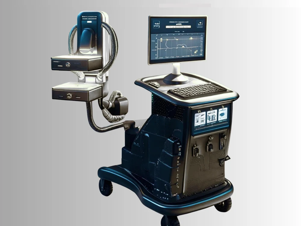
Two small detector heads a few centimetres apart with the breast lightly compressed between them. The patient sits at the machine and each view takes about ten minutes. Because the detectors are close to the tissue, the resolution in the plane of the image is about a millimetre and a half, roughly three times better than a whole-body scanner manages.
Also called: Positron Emission Mammography. Arabic: تصوير الثدي بالإصدار البوزيتروني. Device Lab 6 of 69 in BMEssentia Medical City, 2 configurations.
Overview: the configurations
Opposed-planar PEM scanner
Two small detector heads a few centimetres apart with the breast lightly compressed between them. The patient sits at the machine and each view takes about ten minutes. Because the detectors are close to the tissue, the resolution in the plane of the image is about a millimetre and a half, roughly three times better than a whole-body scanner manages.
Dedicated breast PET ring
A complete detector ring, with the patient lying prone and the breast hanging through an aperture into it. No compression at all. Lines of response arrive from every direction, so the volume reconstructs properly in three dimensions rather than as two flat views.
How it works: the main parts
What each part does, from the PEM lessons in the 3D Device Lab.
- Scintillator crystal array
- A block of small transparent bars, each about two millimetres square and a centimetre or so deep, packed side by side behind the face of every detector. This is the only part of the machine that a 511 keV photon ever touches, and everything after it is dealing with the consequences.
- Photosensor array
- The layer directly behind the crystal that turns its flashes of light into electrical pulses. On a modern breast system these are solid-state devices a few millimetres square rather than the glass photomultiplier tubes that older scanners used.
- Front-end electronics
- The boards immediately behind the photosensors, one set per detector block. They take a small fast charge pulse and turn it into two numbers: how much energy, and at what time.
- Coincidence processor
- A single board or module, usually inside the cart rather than in the detector, that receives every individual detected event from every block and decides which of them belong together in pairs.
- Out-of-field shielding
- Lead or tungsten plate built into the sides and back of the detector assembly, and often a shaped shield between the detectors and the patient’s torso. It is heavy, it is invisible from outside, and it is a large part of why the assembly is as massive as it is.
- Reconstruction workstation
- A computer inside the cart, with more cooling than its size suggests, that turns a list of several tens of millions of lines of response into the images on the review screen.
- Review display
- The screen the operator and the reporting radiologist actually look at, mounted on an arm above the cart, showing either a page of small plane images or a set of orthogonal views depending on which configuration produced the study.
- Keyboard and operator console
- A keyboard, a pointing device and a small set of dedicated controls on the cart top, from which the study is set up, the patient identified and the acquisition started and stopped.
Explore it in the Device Lab
- Two photons, back to back, and what follows from it: A positron is emitted, travels a short distance losing energy, and annihilates with an electron. The rest mass of both becomes two photons of 511 keV each, and because momentum has to balance they leave in opposite directions.
- What the sharpness is actually made of: The resolution figure quoted for a system is not one thing. It is four independent blurs combined in quadrature and then multiplied by a factor for the reconstruction itself.
- The one line that separates the two machines: Take the in-plane resolution of a system and divide it by the sine of half its angular coverage. That gives the resolution along the axis it does not watch.
- Why more tracer stops helping: True coincidences rise in proportion to the activity. Random coincidences, two unrelated events landing inside the same timing window and being paired, rise as its square, because either one of two independent rates can supply either half of the pair.
- The lesion that fades instead of vanishing: A lesion smaller than the resolution does not disappear. It is smeared across a larger volume, so the total counts belonging to it are preserved while the peak value falls. What is left of its true contrast is the recovery coefficient.
Common faults and what to check
Most reported device problems (FDA MAUDE)
| Device problem | Reports |
|---|---|
| Nonstandard Device | 89 |
| Unintended System Motion | 81 |
| Loose or Intermittent Connection | 74 |
| Component Falling | 73 |
| Device Operates Differently Than Expected | 56 |
| Mechanical Problem | 53 |
Raw counts of adverse event reports for this device type (FDA product code KPS), all years. Not adjusted for the number of units in use, so they show what gets reported, not failure rates. Report text is never copied.
Planned maintenance checks
From the BMEssentia procedure library: every 12 months, about 120 minutes, electrical class I, applied part type B.
| Check | Pass when |
|---|---|
| Detector heads and paddles Inspect the detectors, paddles and the compression mechanism. | No damage. |
| Electrical safety Test to IEC 62353. | Within limits. |
| QC and compression Run the manufacturer's QC; check compression force display. | Within limits. |
Test instruments: Manufacturer's QC source and phantom.
Recent FDA recalls
| Recall | Product and reason |
|---|---|
| Z-2229-2008 2008-09-11 | Naviscan PET Systems: Naviscan PET Systems, Inc ("Naviscan") PEM Flex Solo II PET Scanner. The PEM Flex Solo II images small body parts with This field correction is being initiated as a result of the firm's investigation into a report from a user indicating that the motorized compression exceeded 25 pounds of compression force during the pre-scan positioning. Root cause: Device Design. |
| Z-0041-2008 2007-10-11 | Naviscan PET Systems: PEMView Software used with Naviscan PEM Flex Solo I and PEM Flex Solo II PET Scanner, for nuclear medicine imaging. Naviscan has identified a potential safety issue with all versions of PEM View Software commonly used for analyzing patient images acquired on the PEMFIex product line. Root cause: Software design. |
From the FDA recall database (openFDA), newest first, imported 2026-10-22. 2 recalls on record for this device type. Recall text is the FDA's public text; each recall links to the official record.
Learn it, see it in 3D, ask about it
- Ask Essentia about the PEM: answers from named sources, with the device open in its 3D Device Lab beside the answer.
- The PEM on the BMEssentia Platform: the Device Library course, 13 sections from physics to maintenance.
- BMEssentia Medical City: Device Lab 6, the PEM fully assembled in 3D, with lab tasks and the simulations.
Previous: SPECT. Next: Cardiopulmonary Bypass. All 69 devices.
Ask about the PEM
Sign in with your BMEssentia account, or create one, and ask anything about this device.
Ask Essentia