

Device Lab 3
PET/CT
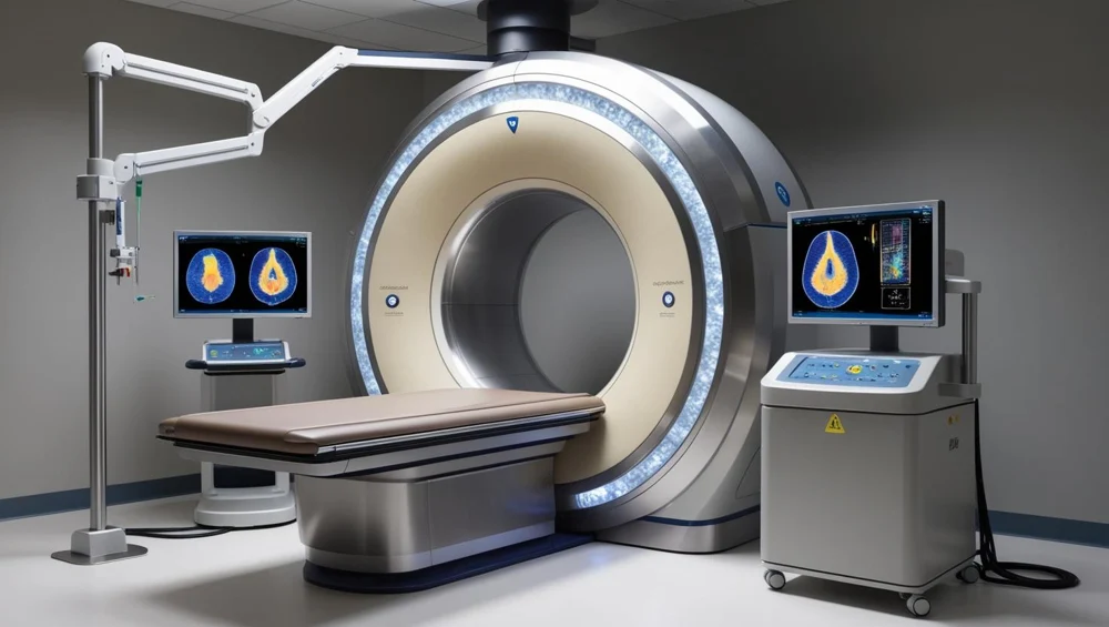
The scanner housing joins two measurement systems around one patient pathway. The front station contains CT; the PET ring sits behind it.
Also called: Pet Scanner, Positron Emission Tomography. Arabic: التصوير المقطعي بالإصدار البوزيتروني. Device Lab 3 of 69 in BMEssentia Medical City, 2 configurations.
How it works: the main parts
What each part does, from the PET/CT lessons in the 3D Device Lab.
- CT acquisition assembly
- CT measures how an external X-ray beam is attenuated through the patient. Its source and opposing detector array rotate around the bore.
- PET detector ring
- The PET ring surrounds the patient with many detector modules. Each module contributes measurements; no single block produces the final image.
- Scintillator crystals
- The scintillator is where deposited gamma energy becomes light. A PET photon can interact in the crystal and produce a short flash too small and fast to see directly.
- Silicon photomultipliers
- The silicon photomultiplier converts the scintillator flash into an electrical signal. It contains many tiny avalanche cells that respond to incoming light.
- Timing and energy readout
- Readout electronics turn a pulse into estimates of energy, arrival time and channel location. Those estimates decide whether an event is useful.
- Coincidence processor
- The coincidence processor compares detections and accepts pairs that satisfy the acquisition conditions. A pair can be consistent with one annihilation, but timing agreement alone cannot prove that both photons came from the same event.
- Patient support and drive
- The patient support establishes the moving coordinate system shared by PET and CT. The narrow top, drive and position feedback work together.
- Positioning controls
- The gantry controls let the technologist position the support while watching the patient. Their location matters: a control is useful only when the operator can see the movement and reach the relevant stop or adjustment.
Explore it in the Device Lab
- When do two detections become a pair?: The exercise compares a fixed teaching event set under two timing windows. Widening the window admits more pairs, including unrelated ones. Predict the trade-off, change the window, then run and pause the illustration.
- A detector sector loses response: A synthetic uniform object should produce a consistent response under controlled conditions. Reduce a detector sector response and watch the illustrative image develop a deficit. This is a teaching response model, not a reconstruction algorithm or a calibrated scanner.
- Two datasets, one patient position: The synthetic emission pattern and anatomical outline start aligned. Move the reference outline and observe where the overlay no longer agrees. Real CT-based attenuation correction also depends on the relationship between these datasets.
- Axial coverage changes the sampling: Compare a shorter axial ring with the longer architecture. The latter surrounds more of the patient axis at once. The mechanism displays a coverage band whose length follows the selected architecture.
Common faults and what to check
Guided fault cases in the Device Lab
- A repeatable detector-sector deficit: A uniform-phantom result shows a sector deficit. A patient examination is waiting.
- The PET and CT maps disagree: The fused synthetic phantom has a spatial mismatch.
- Cooling hold during acquisition: A cooling condition stops the teaching acquisition.
- Local image, missing archive study: Local reconstruction is complete, but archive transfer failed.
Training cases written by BMEssentia for teaching.
Most reported device problems (FDA MAUDE)
| Device problem | Reports |
|---|---|
| Nonstandard Device | 89 |
| Unintended System Motion | 81 |
| Loose or Intermittent Connection | 74 |
| Component Falling | 73 |
| Device Operates Differently Than Expected | 56 |
| Mechanical Problem | 53 |
Raw counts of adverse event reports for this device type (FDA product code KPS), all years. Not adjusted for the number of units in use, so they show what gets reported, not failure rates. Report text is never copied.
Planned maintenance checks
From the BMEssentia procedure library: every 12 months, about 240 minutes, electrical class I, applied part type B.
CT part as for the CT scanner. PET quality control follows the manufacturer and NEMA NU 2 at acceptance; daily QC uses the manufacturer's source.
| Check | Pass when |
|---|---|
| Gantry, table and room Inspect the gantry, table, lasers, the hot lab shielding and warning signs. | No damage; signs in place. |
| Electrical safety Test table and accessories to IEC 62353. | Within limits. |
| PET QC and SUV Review daily QC trends; scan the uniform phantom and compare SUV. | Within the manufacturer's limits and SUV tolerance. |
| Registration and CT Check PET/CT registration with the manufacturer's test; CT checks as for the CT scanner. | Within tolerance. |
Test instruments: Manufacturer's daily QC source; NEMA NU 2 phantom (acceptance and annual, physics); Dose calibrator (radiopharmacy).
Recent FDA recalls
| Recall | Product and reason |
|---|---|
| Z-3283-2026 2026-09-25 | Shanghai United Imaging Healthcare Co., Ltd.: Positron Emission Tomography and Computed Tomography System, Model: uMI 550 with software Positron emission tomography and computed tomography x-ray systems have a software problem when Split or Local function is enabled, and multiple RIS exams are merged into a single study, that prevents patient images from. Root cause: Software design. |
| Z-3284-2026 2026-09-25 | Shanghai United Imaging Healthcare Co., Ltd.: Positron Emission Tomography and Computed Tomography System, Model: uMI Panorama 35 with software Positron emission tomography and computed tomography x-ray systems have a software problem when Split or Local function is enabled, and multiple RIS exams are merged into a single study, that prevents patient images from. Root cause: Software design. |
| Z-3285-2026 2026-09-25 | Shanghai United Imaging Healthcare Co., Ltd.: Positron Emission Tomography and Computed Tomography System, Model: uMI Panorama GS with software Positron emission tomography and computed tomography x-ray systems have a software problem when Split or Local function is enabled, and multiple RIS exams are merged into a single study, that prevents patient images from. Root cause: Software design. |
| Z-3286-2026 2026-09-25 | Shanghai United Imaging Healthcare Co., Ltd.: Positron Emission Tomography and Computed Tomography System, Model: uMI Panvivo with software Positron emission tomography and computed tomography x-ray systems have a software problem when Split or Local function is enabled, and multiple RIS exams are merged into a single study, that prevents patient images from. Root cause: Software design. |
| Z-3287-2026 2026-09-25 | Shanghai United Imaging Healthcare Co., Ltd.: Positron Emission Tomography and Computed Tomography System, Model: uMI Panvivo S with software Positron emission tomography and computed tomography x-ray systems have a software problem when Split or Local function is enabled, and multiple RIS exams are merged into a single study, that prevents patient images from. Root cause: Software design. |
From the FDA recall database (openFDA), newest first, imported 2026-10-22. 208 recalls on record for this device type. Recall text is the FDA's public text; each recall links to the official record.
Learn it, see it in 3D, ask about it
- Ask Essentia about the PET/CT: answers from named sources, with the device open in its 3D Device Lab beside the answer.
- The PET/CT on the BMEssentia Platform: the Device Library course, 13 sections from physics to maintenance.
- BMEssentia Medical City: Device Lab 3, the PET/CT fully assembled in 3D, with lab tasks and the simulations.
Previous: MRI. Next: CT Scanner. All 69 devices.
Ask about the PET/CT
Sign in with your BMEssentia account, or create one, and ask anything about this device.
Ask Essentia