

Essentia / Devices / Endoscope
Device Lab 34
Endoscope
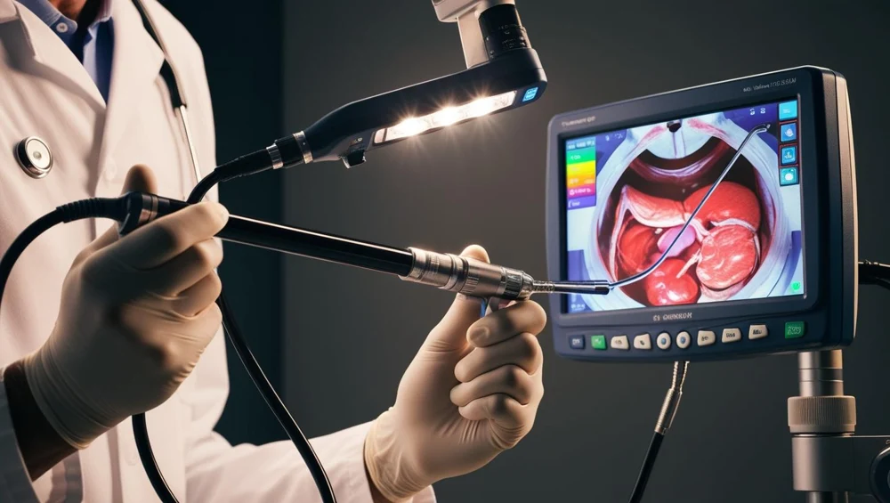
A coherent bundle of some twenty-three thousand fibres carries the image up the insertion tube to an eyepiece, and two incoherent bundles carry light the other way from a lamp in the stack. The objective forms a real image on the distal face of the image guide, every fibre relays one picture element of it, and what a person looks at is the proximal face of the same bundle.
Also called: Gastroscope, Flexible Endoscope. Arabic: المنظار الداخلي. Device Lab 34 of 69 in BMEssentia Medical City, 2 configurations.
Overview: the configurations
Fibre-optic image guide, eyepiece and light bundles
A coherent bundle of some twenty-three thousand fibres carries the image up the insertion tube to an eyepiece, and two incoherent bundles carry light the other way from a lamp in the stack. The objective forms a real image on the distal face of the image guide, every fibre relays one picture element of it, and what a person looks at is the proximal face of the same bundle.
Chip-on-tip sensor and distal light-emitting diode
The objective forms its image directly on a sensor three millimetres behind it, inside the distal tip. What travels up the insertion tube is a signal on a cable six tenths of a millimetre across, and the light comes from a diode at the tip rather than from a lamp in the stack, so no glass runs the length of the scope at all.
How it works: the main parts
What each part does, from the Endoscope lessons in the 3D Device Lab.
- Distal tip and its four apertures
- A black moulded cylinder about ten millimetres across and fourteen long, at the end of a metre of dark flexible tube. Look at it end-on and there are four openings in the face, each a different size, and one of them has a tiny bent spout beside it.
- Objective lens assembly and the field of view
- Three glass elements smaller than a grain of sugar with an aperture stop between them, sitting behind the window in the distal face. The whole assembly is about five millimetres long and there is no focus control anywhere on the instrument.
- Bending section, articulation rings and outer sheath
- The last ten centimetres before the tip, and it is obviously different from the rest of the shaft: softer, more willing, and it stays where it is put. Squeeze it and you can feel discrete rings under the rubber.
- Angulation wires, coil pipes and the control knobs
- Two knobs on the side of the control body, one inside the other. The large one is up and down, the small one is left and right, and each has a lever beside it that locks it.
- Insertion tube: helical band, braid and sheath
- A metre of dark, slightly matt tube about the thickness of a finger, marked with pale rings every five centimetres. It feels almost incompressible along its length and quite willing to curve.
- Biopsy and suction channel
- A pale fluoropolymer tube running the length of the instrument, two and a half to three and a half millimetres across, from a port on the control body to the largest opening in the distal face. On the instrument you never see it.
- Instrument port, biopsy valve and the suction branch
- A short boss angled forward off the top of the control body with a soft rubber cap in it. The cap has a slit in it.
- Air and water channels, nozzle and valve
- A blue-capped button on the control body with a hole in the top of it, and two narrow lumens under a millimetre across running the whole length of the instrument and the cord. Leave the button alone and nothing happens.
Explore it in the Device Lab
- The exponent that decides everything: Put every lumen in one shaft side by side and compare their resistances, then feed them all from one fitting and watch where the delivery goes.
- Why a flush carries rather than scrubs: Raise the flush rate on the working channel until the flow stops being laminar, and then try the same thing on a lumen under a millimetre across.
- Where the image is formed decides how wide the channel is: Add up the cross-sectional area of everything the image path occupies on each configuration, take the difference, and spend it on the working channel.
- A hole nobody can see: Pressurise the interior to a couple of hundred millimetres of mercury, submerge the instrument, and angulate it fully while watching for bubbles. Then work out how small a hole that finds.
- The elegant half, and why it is not the problem: Compute the acceptance angle from two refractive indices, the packing fraction from a core and a pitch, the resolution from a sampling grid, and the depth of field from the same grid. Then note that not one of them is a failure mode.
Common faults and what to check
Most reported device problems (FDA MAUDE)
| Device problem | Reports |
|---|---|
| Failure to Clean Adequately | 23,740 |
| Break | 14,069 |
| Poor Quality Image | 12,205 |
| No Display/Image | 12,085 |
| Replace | 10,356 |
| Communication or Transmission Problem | 8,388 |
Raw counts of adverse event reports for this device type (FDA product code FDS, GCJ), all years. Not adjusted for the number of units in use, so they show what gets reported, not failure rates. Report text is never copied.
Planned maintenance checks
From the BMEssentia procedure library: every 12 months, about 60 minutes, electrical class I, applied part type BF.
Scope repair is the manufacturer's. Reprocessing follows HTM 01-06 / ISO 15883 in the decontamination unit.
| Check | Pass when |
|---|---|
| Insertion tube, bending section and distal tip Inspect for bites, kinks, crushed or peeling insertion tube, a damaged bending rubber and a scratched lens. | No damage; bending rubber intact. |
| Leak test Pressurise per the manufacturer and flex the scope while immersed. | No continuous bubble stream. |
| Angulation and channels Check angulation in every direction and that suction and instrument channels pass a brush freely. | Full angulation; channels clear. |
| Electrical safety of the stack Test the processor, light source and monitor as a medical system to IEC 62353. | PE ≤ 0.30 Ω; equipment leakage ≤ 500 µA; applied part BF ≤ 5000 µA. |
Test instruments: Leak tester (manual or the washer's); Electrical safety analyser; Light meter (optional).
Standards engineers test against
IEC 60601-2-18: endoscopic equipment
The particular standard for endoscopes and their light sources and processors.
ISO 15883: washer-disinfectors
The series for washer-disinfectors: part 1 general requirements, part 2 for surgical instruments, part 4 for flexible endoscopes.
Recent FDA recalls
| Recall | Product and reason |
|---|---|
| Z-2761-2026 2026-07-20 | Olympus Corporation of the Americas: Olympus 4K UHD LCD Monitor. Model Number: OEV321UH. Potential for the HDMI and DisplayPort (DP) connectors can become faulty resulting in image loss or image abnormalities. Root cause: Nonconforming Material/Component. |
| Z-1010-2026 2026-01-02 | Applied Medical Resources Corp: Kii Low Profile, Bladed Dual Pack , Qty. 1 5x55mm There is the potential for extended length of the blade obturator after assembly into the cannula and seal housing. Root cause: Nonconforming Material/Component. |
| Z-0309-2026 2025-10-24 | Aesculap Inc: Brand Name: Aesculap It was determined there is the potential of silicone fragments detaching from the yellow sealing component. Root cause: Under Investigation by firm. |
| Z-0310-2026 2025-10-24 | Aesculap Inc: Brand Name: Aesculap It was determined there is the potential of silicone fragments detaching from the yellow sealing component. Root cause: Under Investigation by firm. |
| Z-0311-2026 2025-10-24 | Aesculap Inc: Brand Name: AESCULAP It was determined there is the potential of silicone fragments detaching from the yellow sealing component. Root cause: Under Investigation by firm. |
From the FDA recall database (openFDA), newest first, imported 2026-10-22. 286 recalls on record for this device type. Recall text is the FDA's public text; each recall links to the official record.
Learn it, see it in 3D, ask about it
- Ask Essentia about the Endoscope: answers from named sources, with the device open in its 3D Device Lab beside the answer.
- The Endoscope on the BMEssentia Platform: the Device Library course, 13 sections from physics to maintenance.
- BMEssentia Medical City: Device Lab 34, the Endoscope fully assembled in 3D, with lab tasks and the simulations.
Previous: Retinal Laser. Next: Colonoscope. All 69 devices.
Ask about the Endoscope
Sign in with your BMEssentia account, or create one, and ask anything about this device.
Ask Essentia