

Essentia / Devices / DEXA Scanner
Device Lab 46
DEXA Scanner
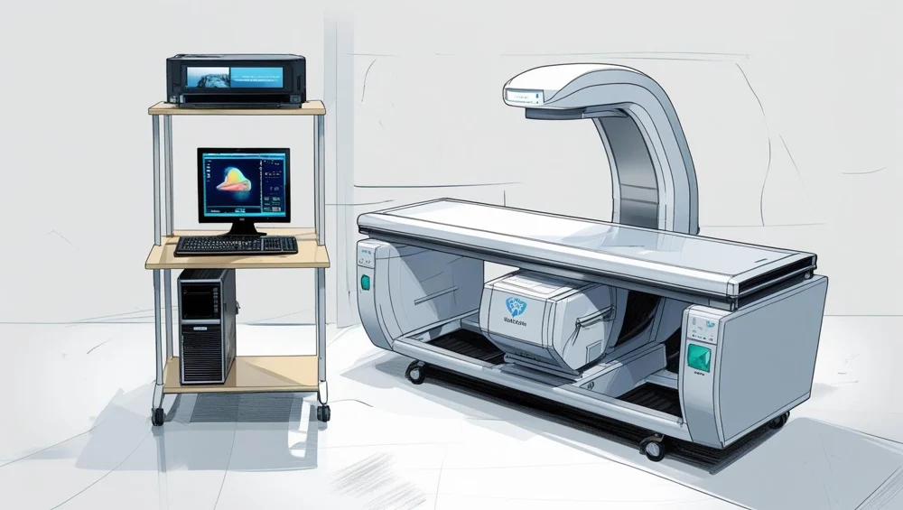
A low table with a C-arm inside it: the tube in the base below the tabletop, a single detector on an arm above the patient, and the two joined so they move as one object. The arm travels the length of the table and the detector head travels across it, so the beam visits the region point by point on a raster. A circular aperture a millimetre across defines the beam.
Also called: Dxa, Bone Densitometer, Bone Density Scanner. Arabic: جهاز قياس كثافة العظام. Device Lab 46 of 69 in BMEssentia Medical City, 2 configurations.
Overview: the configurations
Pencil-beam raster scanner with a single detector
A low table with a C-arm inside it: the tube in the base below the tabletop, a single detector on an arm above the patient, and the two joined so they move as one object. The arm travels the length of the table and the detector head travels across it, so the beam visits the region point by point on a raster. A circular aperture a millimetre across defines the beam.
Narrow fan-beam scanner with a linear detector array
The same low table and the same C-arm, with a slit instead of a pinhole and a linear array of a hundred and twenty-eight elements instead of one detector. The arm travels the length of the table once and the fan covers the width, so the region is acquired in a single sweep. The array carries its own channel gain and geometric calibration, because a hundred and twenty-eight detectors that do not agree with each other draw stripes.
How it works: the main parts
What each part does, from the DEXA Scanner lessons in the 3D Device Lab.
- X-ray tube, stationary anode and oil bath
- A small cylinder lying on its side in the bottom of the table, under the patient, with its port facing upwards. It is a fraction of the size of the tube in a radiography room and it runs at a couple of milliamperes rather than hundreds, because a densitometry scan is minutes of continuous output rather than a flash of milliseconds.
- Switched high-tension supply and potential control
- A small oil-filled tank and two circuit boards in the base of the table, well under a kilowatt in total. The part that makes this a dual-energy machine rather than an X-ray machine is one of those boards: it alternates the accelerating potential between two values, many times a second, for the whole length of the scan.
- Beam filtration and energy-separation assembly
- A metal disc a hundred and ten millimetres across sitting directly over the tube port, turning in step with the potential switch. Four stations are cut into it: a light absorber for the low pulse, a much heavier one for the high pulse, a reference absorber of known attenuation, and one station left open.
- Low-attenuation scanning tabletop and pad
- A flat panel a little over half a metre above the floor with a thin pad on it, both of them directly in the beam on every single measurement. The table is low because patients who come for this examination are often elderly and have to get onto it unaided, and the panel is carbon-fibre composite rather than the aluminium a radiographic table can afford.
- Positioning aids: knee block, foot separators and arm rests
- Three foam blocks and a pair of radiolucent supports on a rack on the side of the machine. They look like the least technical objects on the device and they are worth more to the precision of the result than any electronic component on it.
- C-arm scanning carriage and longitudinal drive
- A rigid C lying on its side inside and above the table, with the tube in its lower limb below the tabletop and the detector in its upper limb above the patient. The whole C travels the length of the table on a rail, driven by a belt and a motor, and the fact that it is one object rather than two is the most important thing about it.
- Position encoders and the scan-step datum
- A fine scale down the length of the base with a read head on the carriage, a datum flag near one end, and on the raster machine a second encoder on the screw that drives the detector head across the table. Small, cheap, and on one of the two configurations it is the component that supplies the denominator of the answer.
- Detector signal chain and energy discrimination
- A small crate of boards behind the column: a preamplifier, a shaping stage, a discriminator and a counter, in that order. What it produces is not a current and not an image.
Explore it in the Device Lab
- The determinant, and what happens as it shrinks: Harden the high-energy pulse and watch four numbers move together: the two effective energies, the bone-to-soft ratio at each of them, the determinant, and the standard deviation of the areal density at a fixed number of counted photons.
- Where the independence actually comes from: Compute the determinant twice: once with the real attenuation coefficients, and once with only the Compton part of each of them.
- The third unknown there is no equation for: Set the fat fraction the analysis assumes and the fat fraction the patient actually has to different values, and watch the areal bone density move while nothing about the bone changes.
- A density that is not a density: Take one bone, scale it up by a fifth in every dimension without changing what it is made of, and watch what the machine reports.
- The smallest change that means anything: Set the counts per measurement, read the counting floor the physics gives, then set the department’s own measured precision error beside it and read the least significant change.
Common faults and what to check
Most reported device problems (FDA MAUDE)
| Device problem | Reports |
|---|---|
| Device Displays Incorrect Message | 10 |
| Poor Quality Image | 8 |
| Device Stops Intermittently | 5 |
| Unintended Electrical Shock | 3 |
| Device Remains Activated | 2 |
| Incorrect Or Inadequate Test Results | 2 |
Raw counts of adverse event reports for this device type (FDA product code KGI), all years. Not adjusted for the number of units in use, so they show what gets reported, not failure rates. Report text is never copied.
Planned maintenance checks
From the BMEssentia procedure library: every 12 months, about 60 minutes, electrical class I, applied part type B.
| Check | Pass when |
|---|---|
| Arm, table and controls Inspect the C-arm, the table, the emergency stop and the warning light. | No damage. |
| Electrical safety Test to IEC 62353. | Within limits. |
| Phantom QC trend Review the QC chart over the year and scan the phantom. | Within control limits; no drift. |
Test instruments: Manufacturer's spine phantom; Radiation survey meter.
Recent FDA recalls
| Recall | Product and reason |
|---|---|
| Z-1250-2025 2025-02-28 | Medimaps Group: Brand Name: TBS Osteo Potential variability in calculations from fast array scans compared to array scans when operating on Hologic Horizon machines. Root cause: Software Design Change. |
| Z-2373-2024 2024-07-16 | Hologic Inc: Horizon X-Ray Bone Densitometer (DXA) Non-conformance pertains to electromagnetic compatibility requirements according to the international technical standard IEC 60601-1-2 for the safety and essential performance of medical electrical equipment, where the. Root cause: Under Investigation by firm. |
| Z-1709-2020 2020-04-24 | GE Healthcare, LLC: Lunar iDXA bone densitometer An issue with the Lunar iDXA control panel may cause the start button to not release and potentially contribute to an accidental radiation occurrence. Root cause: Radiation Control for Health and Safety Act. |
| Z-1710-2020 2020-04-24 | GE Healthcare, LLC: Lunar iDXA Advance (iDXA with enCORE v16) bone densitometer An issue with the Lunar iDXA control panel may cause the start button to not release and potentially contribute to an accidental radiation occurrence. Root cause: Radiation Control for Health and Safety Act. |
| Z-1711-2020 2020-04-24 | GE Healthcare, LLC: Lunar iDXA Pro/Lunar iDXA Advance (iDXA with enCORE v17) bone densitometer An issue with the Lunar iDXA control panel may cause the start button to not release and potentially contribute to an accidental radiation occurrence. Root cause: Radiation Control for Health and Safety Act. |
From the FDA recall database (openFDA), newest first, imported 2026-10-22. 26 recalls on record for this device type. Recall text is the FDA's public text; each recall links to the official record.
Learn it, see it in 3D, ask about it
- Ask Essentia about the DEXA Scanner: answers from named sources, with the device open in its 3D Device Lab beside the answer.
- The DEXA Scanner on the BMEssentia Platform: the Device Library course, 13 sections from physics to maintenance.
- BMEssentia Medical City: Device Lab 46, the DEXA Scanner fully assembled in 3D, with lab tasks and the simulations.
Previous: Blood Gas Analyser. Next: Fetal Monitor. All 69 devices.
Ask about the DEXA Scanner
Sign in with your BMEssentia account, or create one, and ask anything about this device.
Ask Essentia