

Device Lab 39
ECG
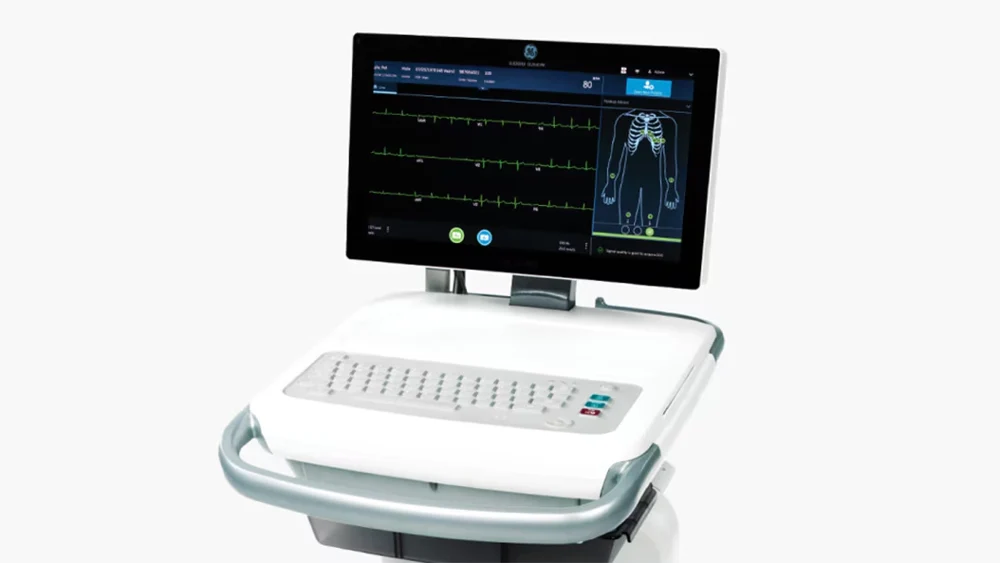
Ten electrodes: one on each limb and six across the chest, every one of them found from bony landmarks by whoever is recording. The four limb electrodes go at the wrists and ankles. Nine of the ten become node potentials and the tenth appears in no lead definition at all, so twelve traces are computed from eight independent measurements.
Also called: Ekg, Electrocardiograph, 12-Lead. Arabic: جهاز تخطيط القلب الكهربائي. Device Lab 39 of 69 in BMEssentia Medical City, 2 configurations.
Overview: the configurations
Twelve-lead diagnostic recorder, ten electrodes
Ten electrodes: one on each limb and six across the chest, every one of them found from bony landmarks by whoever is recording. The four limb electrodes go at the wrists and ankles. Nine of the ten become node potentials and the tenth appears in no lead definition at all, so twelve traces are computed from eight independent measurements.
Continuous five-electrode monitoring configuration
Five electrodes, all of them on the torso: the four limb electrodes moved off the limbs on to the trunk so that an arm can be moved without moving a trace, and one precordial electrode. Four of the five become node potentials and the fifth appears in no lead definition, so seven traces are computed from three independent measurements. The band runs from half a hertz to forty, sampled at two hundred and fifty, and the electrodes stay where they were put for hours or days.
How it works: the main parts
What each part does, from the ECG lessons in the 3D Device Lab.
- Disposable pre-gelled electrode, its element and its contact
- Ten small foam discs on a peel-off strip, each a few centimetres across, each with a metal stud moulded through its back and a bead of wet gel in a shallow well on its front. Tilt the strip and the gel moves.
- Lead wires, the colour code and the yoke
- Ten slim coloured wires, each ending in a clip or a stud socket, each printed with a position name, gathered at a moulded yoke where they all leave for the trunk cable. The colours are the only thing distinguishing one from another once they are on a body.
- Patient trunk cable, its screen and its connector
- One thick cable a couple of metres long, a moulded keyed connector at the instrument end and the lead wire yoke at the other, with a drape hook part way along. It is the heaviest flexible thing on the machine and the one most often replaced.
- The right-leg electrode, which appears in no lead at all
- One disc exactly like the other nine, low on the right side, with a wire in the odd colour out. On the front panel it has an indicator of its own next to the others, and on the twelve panes it has nothing.
- Defibrillation protection and input current limiting
- Immediately behind the patient connector, a row of ten identical little networks on the acquisition board, each a resistor and a clamping device, with nothing to adjust and no indicator. In ordinary use none of it does anything at all.
- Acquisition module and the input multiplexer
- A shielded box behind the patient connector with the ten protected inputs entering one face and a digital ribbon leaving the other. There is nothing to adjust on it and a legend on the lid giving the band and the sample rate for each configuration.
- Wilson central terminal network, and the average nobody measured
- Three identical resistors on the acquisition board, sitting side by side, with one end of each going to a limb input and the other ends joined at a single point. A single track leaves that point and goes to every one of the precordial difference stages.
- Differential amplifier and the electrode offset it must survive
- One instrumentation stage per lead on the acquisition board, a row of identical small packages with no adjustment brought out anywhere on the outside of the instrument. There is nothing on the front panel that changes the gain.
Explore it in the Device Lab
- Move the lower corner, watch a feature appear: Hold the illustrative complex and everything else still, and change only the lower corner. Read the baseline displacement immediately after the deflection.
- Integrate the output and find out why the feature had to exist: Change nothing but the length of the window you integrate over, and watch the total area under the filtered output.
- Move the same corner again, and watch the wander instead: Leave the complex alone. Set a baseline drift of an illustrative one millivolt per second, which is a declared bench control rather than a measurement of anybody, and read the steady output at each corner.
- Move the upper corner, watch amplitude leave: Hold the illustrative upstroke at twenty milliseconds and change only the upper corner. Read the height the apex reaches as a fraction of what went in.
- Delete a channel and ask the rank what it cost: Turn off displayed traces one at a time, in any order, and read the rank of what is left.
Common faults and what to check
Most reported device problems (FDA MAUDE)
| Device problem | Reports |
|---|---|
| Wireless Communication Problem | 100 |
| Appropriate Device Problem Term/Code Not Available | 74 |
| Patient Data Problem | 58 |
| Incorrect Measurement | 46 |
| High Readings | 37 |
| Fire | 35 |
Raw counts of adverse event reports for this device type (FDA product code DPS), all years. Not adjusted for the number of units in use, so they show what gets reported, not failure rates. Report text is never copied.
Standards engineers test against
IEC 60601-2-27: ECG monitoring equipment
The particular standard for electrocardiographic monitoring (bedside and central monitors).
Recent FDA recalls
| Recall | Product and reason |
|---|---|
| Z-3012-2026 2026-08-25 | Midmark Corporation: Midmark IQecg Digital ECG, Model/Catalog Number: 4-000-0061, 4-000-0062 A communication anomoly may result in incorrect electrocardiogram waveforms of a patient and an inaccurate beats-per-minute calculation. Root cause: Software design. |
| Z-2955-2026 2026-08-14 | Philips North America Llc: Cardiac Workstation 5000. Product Number: 860439. Device will intermittently provide inaccurate data related to native heart rate. Root cause: Software design. |
| Z-2956-2026 2026-08-14 | Philips North America Llc: Cardiac Workstation 7000. Product Number: 860441. Device will intermittently provide inaccurate data related to native heart rate. Root cause: Software design. |
| Z-2876-2026 2026-08-04 | GE Medical Systems Information Technologies Inc: GE Healthcare MAC VU360, REF# part number 2030361-023 Software issue issue with the MAC VU360 v3.0 system. Root cause: Software design. |
| Z-0231-2026 2025-10-22 | GE Medical Systems, LLC: MAC VU360 Acquisition Trunk Cable and Module Holder, high performance, multichannel resting electrocardiograph, Model If a user incorrectly places the Acquisition Module into the Acquisition Module Holder, it can result in excessive bending and wear of the Acquisition Trunk Cable. Root cause: Device Design. |
From the FDA recall database (openFDA), newest first, imported 2026-10-22. 98 recalls on record for this device type. Recall text is the FDA's public text; each recall links to the official record.
Learn it, see it in 3D, ask about it
- Ask Essentia about the ECG: answers from named sources, with the device open in its 3D Device Lab beside the answer.
- The ECG on the BMEssentia Platform: the Device Library course, 13 sections from physics to maintenance.
- BMEssentia Medical City: Device Lab 39, the ECG fully assembled in 3D, with lab tasks and the simulations.
Previous: Cardiac Monitor. Next: Holter. All 69 devices.
Ask about the ECG
Sign in with your BMEssentia account, or create one, and ask anything about this device.
Ask Essentia